You can use this free image under the Creative Commons Zero (CC0) public domain license. Support our free section by adding a credit line next to the photo in your design. A suggestion is provided under the title.
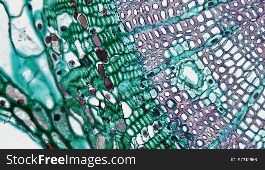
Gymnosperm Stem: Cambium in Two Year Pinus
cross section: Pinus stem magnification: 400x During the first year of growth the cutinized epidermis is replaced by protective growth of cork rich periderm. The outer periderm consists of layers of cork cells, the phellem, which produces waterproofing suberin. Cork cells are dead at maturity. Deep to the phellem is a living layer of cork cambium or phellogen and beneath that, layers of cork parenchyma or phelloderm. Many cells in the periderm contain dark staining tannins. The cortex is divided into a thin outer hypodermis of lignified sclerenchyma cells and thicker inner cortex of thin walled parenchyma cells containing chloroplasts. Large resin ducts are surrounded by secretory parenchyma that produces resins and turpentines. Some cells enlarge into dark staining tyloses. Both endoderm and pericycle are inconspicuous. The vascular cylinder or stele in young stems consists of a ring of vascular bundles interspaced with medullary rays of parenchyma cells. Seasonal activity of the cambium replaces the isolated vascular bundles with well-defined annual rings of secondary phloem and xylem. Xylem is endarch with protoxylem found towards center of the stem and younger metaxylem towards the periphery of the stem. Protoxylem consists of annular and spiral tracheids with only tracheids found in metaxylem. True xylem vessels are lacking. Because of the greater production of xylem, the vascular cylinder is dominated by radially arranged rays of secondary xylem interspaced with medullary rays of parenchyma cells. Conspicuous resin ducts are present throughout the xylem. Phloem is endarch but annual growth the of stem makes it difficult to distinguish between older protophloem to the periphery and younger metapholem towards center of the stem. Phloem lacks companion cells, consisting entirely of sieve tubes and phloem parenchyma. Medullary rays in the secondary phloem include protein rich albuminous cells. A well-defined pith of parenchyma cells is interrupted by a few large resin ducts.
© publicdomainstockphotos | Stock Free Images